Beyond Movement: First Direct Evidence to Support Thinking in the Cerebellum
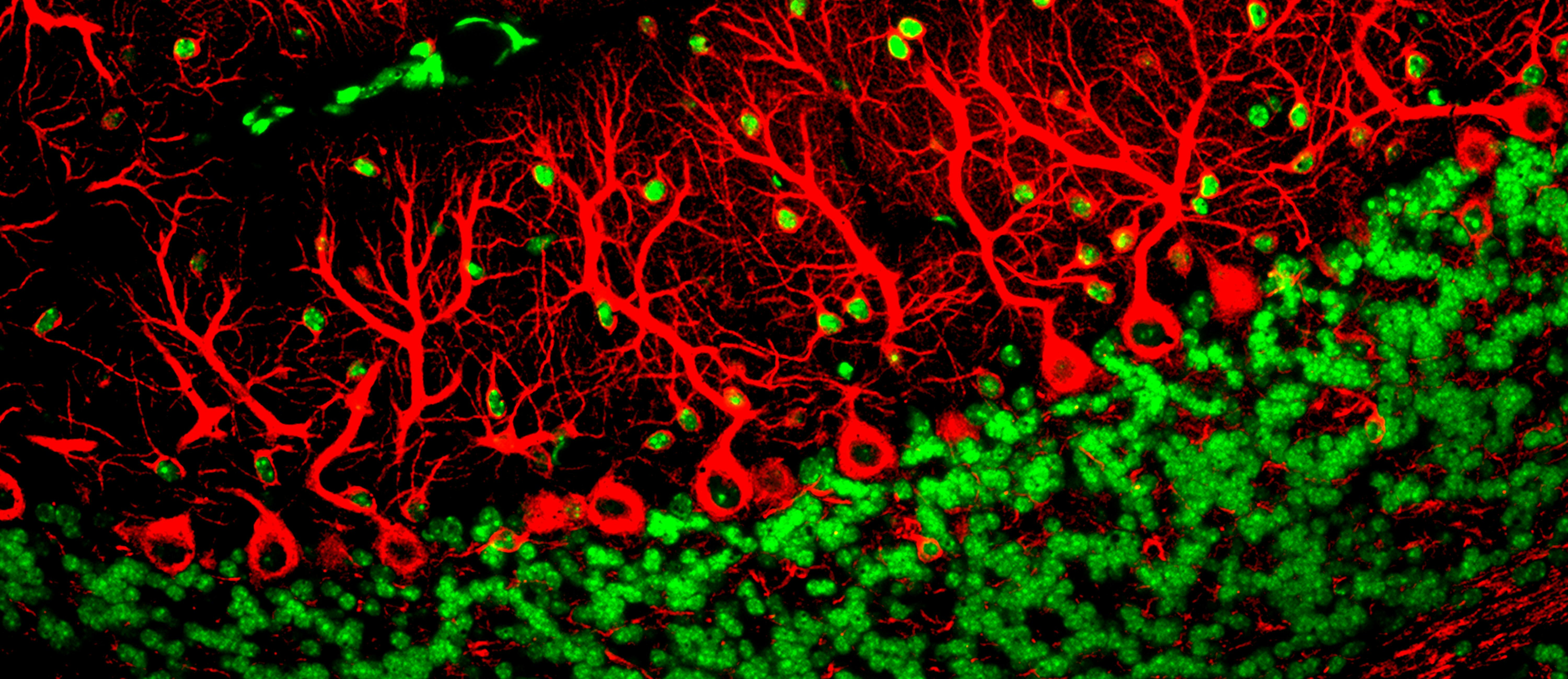
The cerebellum, a striated structure at the back of the brain, is best known for its role in movement. Its densely packed cells (in mammals, the cerebellum houses more than half the brain’s neurons) integrate sensory and internal information to fine-tune movement, helping us learn to hit a baseball or do a backflip.
Past studies, however, have hinted at a broader role for this section of the brain. People with damage to the cerebellum sometimes have trouble with working memory and decision-making. “But aside from lesion studies, there hasn’t been a direct demonstration that this region contributes to cognitive function, which is mostly associated with the frontal cortex,” says Nuo Li, a neuroscientist at Baylor College of Medicine and an investigator with the Simons Collaboration on the Global Brain (SCGB).
Two new studies in mice provide the most direct evidence to date that the cerebellum is involved in short-term memory and decision-making. “The research shows that even tasks that we thought were the job of cortex actually depend a lot on the cerebellum,” says Dora Angelaki, a neuroscientist at New York University and a member of the SCGB’s executive committee who was not involved in the research.
Angelaki says she was not surprised by the findings. The two structures are highly connected, with each part of the cerebellum connected to different parts of the cortex. “No one had shown really the functional significance of that anatomy,” Angelaki says.

The research highlights a growing effort to explore how different regions communicate, a departure from traditional practice in what has been a largely compartmentalized discipline; most neuroscience research to date has focused on individual brain regions, such as the cerebellum or the frontal cortex. “Everyone has a favorite area, and they only record from that area,” Angelaki says.
Coordinated communication
Li and collaborators study short-term memory by training mice to respond to an object that appears near the front or back of their head. After a short delay, the animals indicate the object’s position by licking to the left or the right, earning a water reward. The delay forces the mice to remember the object’s location or their impending choice — and gives scientists the opportunity to study this internal state. “They have to make a decision about the sensory stimulus and use short-term memory to make the correct movement,” Li says.
In previous research, Li and others have shown that neurons in the frontal cortex are active during this delay period, and that the cells’ activity predicts what choice the animal will make. “Presumably this activity in the frontal cortex reflects some kind of short-term memory signal that allows the mouse to know what to do in the future,” Li says. “How does the cerebellum function in this process, and how is it important for maintaining or orchestrating this type of memory activity?”
Li’s team found that, like neurons in the frontal cortex, cerebellar cells are active during the delay period and predict the animal’s subsequent decision. “So memory activity is present both in the frontal cortex and the cerebellum,” Li says.
Moreover, these cerebellar neurons are essential for making the correct choice. When researchers used optogenetics to silence them during the delay period, they found that animals can still make the desired movement — licking left or right — but they no longer choose the appropriate direction. “They behave as if they forgot what to do,” Li says.

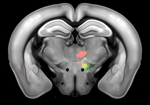
To assess how the cerebellum and frontal cortex interact, researchers silenced one area while recording from the other. Silencing one region erases the other’s ability to predict the animal’s subsequent choice. “It seems these two regions are not chattering independently,” Li says. “They are active in a coordinated way, where one is talking with the other, even when the body is not in motion.” The research was published in Nature in October.
A second study, published in eLife in August, found that the cerebellum is also essential for decision-making. In the experiment, mice received puffs of air on their left or right whiskers and had to decide which side received more puffs, licking on that side to receive a reward. The task required the animals to gather information over time to make their decision.
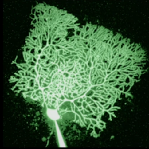
Researchers found that as evidence accumulates, a type of neuron in the cerebellum called Purkinje cells ramped up their firing. The activity of some cells corresponded to a growing number of right-side puffs, while others represented left-side puffs. These cells encoded the animals’ eventual decision. Inactivating the region using a drug called muscimol disrupted the animals’ performance. As in Li’s study, the animals could still lick left or right, but they seemed to be guessing the correct answer — their choices were no better than chance.
“The cerebellum sends signals to the neocortex, including regions known to be important for evidence accumulation, such as the frontal cortex and parietal cortex,” says Sam Wang, a neuroscientist at Princeton University, who led the work. “The region has a signal, it’s necessary, and is sending [the information] to other brain regions involved in the task.”
Motor learning
The findings suggest that the cerebellar circuitry that underlies motor learning might have a broader function. One of the cerebellum’s most iconic functions is its role in cementing new motor skills, such as throwing darts. As we learn, cerebellar circuits compare internal information about an intended movement with external sensory information about the actual movement. The circuits use that information to modify future movements, enhancing the accuracy of subsequent throws, for example.
Wang and Li say their results support the idea that the same error-correcting circuitry that allows the cerebellum to fine-tune movement may also help the brain evaluate evidence in other contexts, such as when we’re choosing whether to hit or hold in a game of blackjack. “It appears it’s playing an analogous role in working memory that it plays in the control of movement,” Wang says.

“The circuitry is set up to perform those kinds of computations — it’s conceivable that a similar kind of computation can take place for activities related to our thoughts rather than to our body,” Li says. “That’s been a provocative hypothesis in the field for some time. Studies like Sam’s and our study says that could be a real possibility.”
Indeed, Wang’s team found neural signals corresponding to moments when the animal made a mistake. “That’s important because the cerebellum is known to learn from errors to guide future actions — it’s a good learning machine,” Wang says. “The fact that it contains error signals in a working-memory task suggests it could learn from those errors.”
Crossing borders
The findings also highlight the importance of studying the interactions between brain regions. Scientists have traditionally thought of the frontal cortex as a sort of command center, Li says. The new results paint a different picture, in which different regions work together in a coordinated way. “Each region probably has some specialty in information processing,” Li says. “It’s through coordination that they can orchestrate a behavior.”
A number of SCGB-funded projects are exploring how different parts of the brain communicate by analyzing the relationship between populations of neurons in multiple areas. Li’s group, along with Shaul Druckmann’s lab at Stanford University, Karel Svoboda’s lab at the Howard Hughes Medical Institute’s Janelia Research Campus, and Xiao-Jing Wang’s group at New York University, will map activity across the brain as mice perform a single behavior. “That’s a starting point to understand where signals reside in the brain and how they are related to each other,” Li says.
“As a field, we are still at the beginning stage of understanding multiregional communication in the brain,” Li says. “For any one behavior, we don’t know comprehensively what regions are involved and how differently regions communicate. That’s an important, exciting direction the field is moving toward.”
Li points out that most researchers studying short-term memory and decision-making don’t include the cerebellum or any other type of multiregional communication. “This, I think, will be a challenge, how to take biology into account when thinking about multiregional models,” he says. “What does it mean to build a multiregional model, and why would you need it?” Druckmann and Wang, both theorists, are beginning to think about this kind of question.


